![]() Figure 2 of
Kanagavalli, Mol Vis 2007;
13:1161-1168.
Figure 2 of
Kanagavalli, Mol Vis 2007;
13:1161-1168.
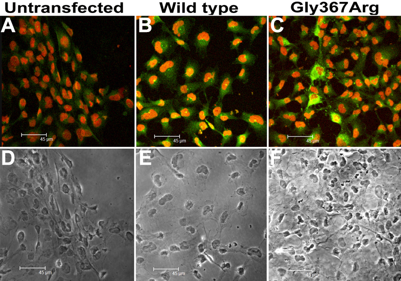
Figure 2. Confocal microscopic image of trabecular meshwork cells transfected with wild type and mutant myocilin
A: Untransfected TM cells showed no endogenous expression of myocilin. B: The TM cells showed wild type myocilin expression in the perinuclear region. C: Intense staining of Gly367Arg mutant myocilin in the perinuclear region of TM cells. D-F: Phase contrast images of A-C, respectively.