![]() Figure 1 of
Kanagavalli, Mol Vis 2007;
13:1161-1168.
Figure 1 of
Kanagavalli, Mol Vis 2007;
13:1161-1168.
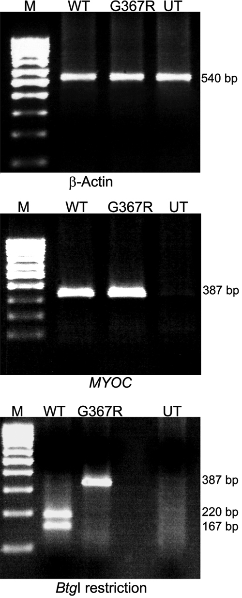
Figure 1. Reverse transcriptase-polymerase chain reaction analysis and restriction digestion
Confirmation of transfection experiment by RT-PCR and BtgI restriction RT-PCR was carried out using exonic primers to amplify the specific region of exon 3 of MYOC which resulted in 387 bp products. The left top panel showed β-actin amplification (540 bp) as an internal control in all three lanes. MYOC amplification was observed in the second and third lanes corresponding to TM cells transfected with wild type and Gly367Arg and was not seen in the control (untransfected). In the third lane of the bottom panel, Gly367Arg caused loss of restriction site for BtgI and confirmed the method of Gly367Arg MYOC transfection in TM cells. The wild type PCR products resulted in two bands of 220 and 167 bp as seen in the second lane. M- Marker; WT-wild type; G367R-Gly367Arg; UT-untransfected.