![]() Figure 2 of
Lu, Mol Vis 2007;
13:1154-1160.
Figure 2 of
Lu, Mol Vis 2007;
13:1154-1160.
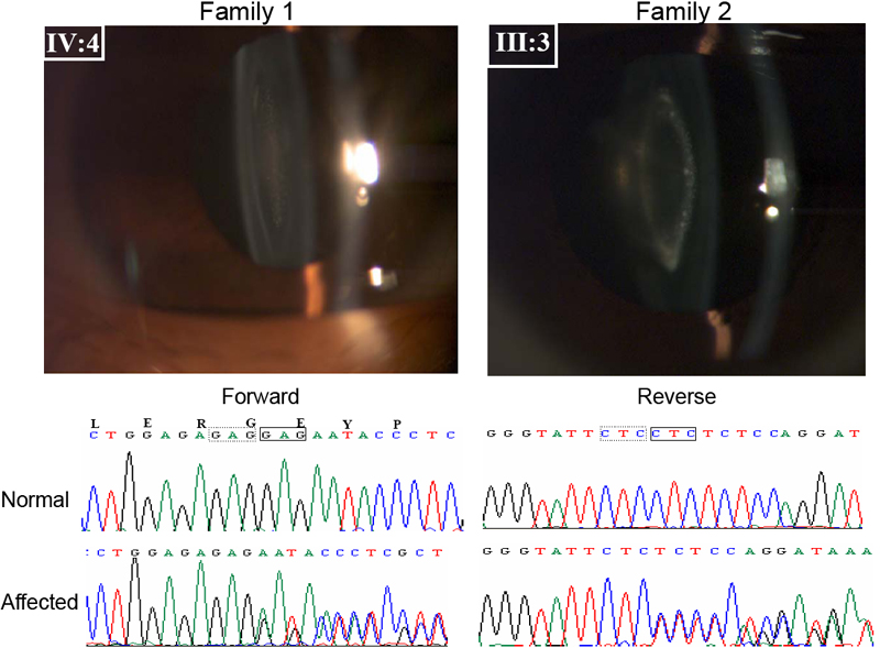
Figure 2. Lens phenotypes and detection of identical CRYBA1 mutations in Families 1 and 2
Shown in the top panel are direct slit-lamp photographs showing typical opacities of pulverulent nuclear cataract in affected member IV:4 in Family 1. Member III:3 of Family 2 shows lamellar pulverulent cataract with a few dot-like opacities restricted to the lamellae together with a transparent normal embryonic nucleus. In the bottom panel are shown deletions of 3 bp in exon 4 of CRYBA1 in the two families as illustrated by the forward and reverse sequences shown, corresponding to Family 1 and Family 2, respectively. These mutations leads to loss of a glycine residue at amino acid position 91 (ΔG91).