![]() Figure 5 of
Mitchell, Mol Vis 2007;
13:1144-1153.
Figure 5 of
Mitchell, Mol Vis 2007;
13:1144-1153.
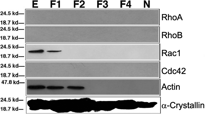
Figure 5. Western blot analysis of the expression patterns of RhoA, RhoB, Rac1, and Cdc42
from four-week-old rat lens as determined by western blot analysis lens epithelium (E), cortical fiber cells (F1), subcortical fiber cells (F2), middle layer of fiber cells (F3), inner layers of fiber cells (F4), and nuclear fiber cells (N) from four-week-old rat lens as determined by western blot analysis. As comparison, the expression patterns of β-actin and α-crystallin were also included. Note that as observed in the mouse lens, Rac1 was expressed mainly in the lens epithelium and to a lesser degree in F1 lens fiber cells. β-Actin was detectable in the lens epithelium, cortical (F1), and sub-cortical (F2) fiber cells. In contrast, α-crystallin is present in both lens epithelium and all layers of lens fiber cells.