![]() Figure 3 of
Mitchell, Mol Vis 2007;
13:1144-1153.
Figure 3 of
Mitchell, Mol Vis 2007;
13:1144-1153.
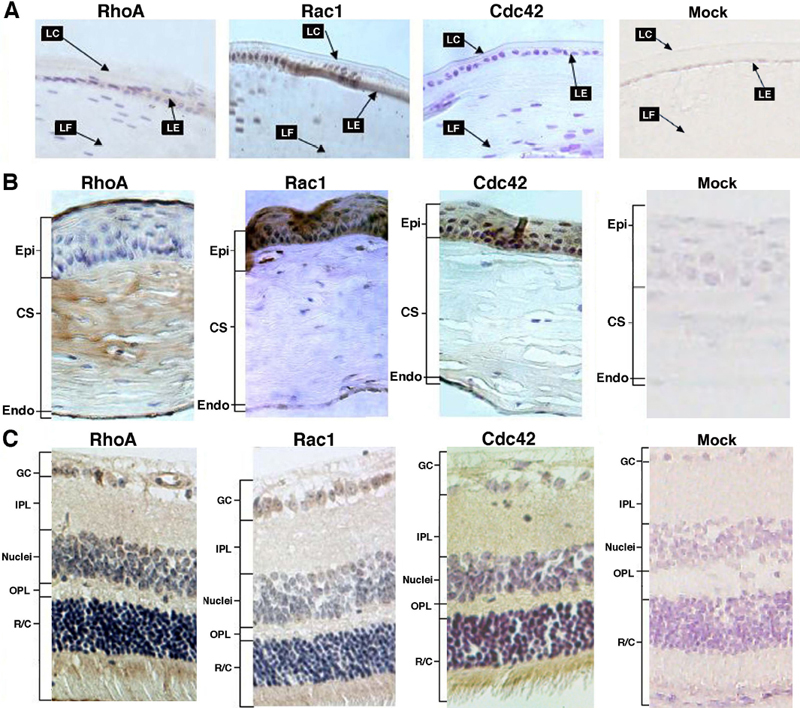
Figure 3. Immunocytochemistry analysis of the expression patterns of RhoA, Rac1 and Cdc42 in adult mouse eye
A: Expression of the three small GTPases in the adult mouse lens was investigated. While Rac1 was detected in both lens epithelium and fiber cells, expression of RhoA and Cdc42 were barely detectable in the adult mouse lens. Compared with the control, the nuclei of both epithelial and fiber cells of mouse lens displayed higher background. LC: lens capsule; LE: lens epithelium; LF: lens fiber cells. B: Expression of the three small GTPases in the adult mouse cornea was followed. Strong RhoA expression was detected in the top layer of corneal epithelial and endothelial cells. Some RhoA was detected in fiberoblast cells. Expressions of Rac1 and Cdc42 were strong in the epithelial layers but relatively lower in the endothelial cell. No obvious staining was seen in the corneal stroma. Epi: corneal epithelial cell layers; CS: corneal stroma; Endo: corneal endothelial cell layers. C: Expression of the three small GTPases in the adult mouse retina was investigated. Both RhoA and Cdc 42 were highly expressed in the photoreceptors, the horizontal/amacrine/Muller cell layers, and in some ganglion cells. In contrast, strong expression of Rac1 was only observed in the photoreceptors. GC: ganglion cells; IPL: inner plexiform; Nuclei: horizontal/amacrine/Muller cell layers; OPL: outer plexiform; R/C: rods and cones (photoreceptors). Mock sections in the rightmost panels of A, B, and C were processed in the same way as the immunocytochemistry results shown for RhoA, Rac1, and Cdc42 in the same figure except that the primary antibodies were replaced with normal rabbit serum.