![]() Figure 1 of
Mitchell, Mol Vis 2007;
13:1144-1153.
Figure 1 of
Mitchell, Mol Vis 2007;
13:1144-1153.
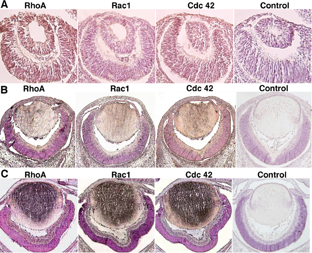
Figure 1. Immunocytochemistry analysis of the expression patterns of RhoA, Rac1, and Cdc42 during mouse eye development
A: Expression of RhoA, Rac1, and Cdc42 on day 11.5 pc mouse embryonic eye. RhoA was highly expressed in the peridermal cornea, lens vesicle and differentiating retina. Compared with RhoA, Cdc42 was expressed in a similar pattern in these compartments but the level was lower. Expression of Rac1 in the three compartments was detectable but in a much lower level in comparison to both RhoA and Cdc42. B: Expression of RhoA, Rac1, and Cdc42 on day 14.5 pc mouse embryonic eye. Expression of the three GTPases is elevated in the differentiating cornea, lens fibers, and the inner region of the neuroblastic layer. C: Expression of RhoA, Rac1, and Cdc42 on day 17.5 pc mouse embryonic eye. Note that strong expressions of RhoA, Rac1, and Cdc42 were observed in the epithelial and primary fiber cells of the embryonic lens. To a less degree, these small GTPases are expressed in the cortical fiber cells, corneal epithelial and endothelial cells, and differentiating retina.