![]() Figure 3 of
Ono, Mol Vis 2007;
13:1138-1143.
Figure 3 of
Ono, Mol Vis 2007;
13:1138-1143.
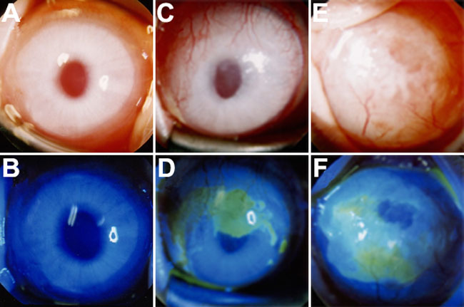
Figure 3. Representative cases from the three groups at two months after surgery
In most of the cases in the Conj-AM group, the cornea was clear with minimal neovascularization and had no epithelial defects (A and B). In the AM Alone group, moderate neovascularization and opacity was present in the midperipheral to peripheral cornea and epithelial defects in the central area (C and D). In the No Transplantation group, advanced neovascularization and opacity were present throughout the cornea, and epithelial defects were in the central area (E and F).