![]() Figure 2 of
Ono, Mol Vis 2007;
13:1138-1143.
Figure 2 of
Ono, Mol Vis 2007;
13:1138-1143.
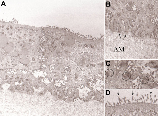
Figure 2. Transmission electron micrographs of rabbit conjunctival epithelial cells grown on amniotic membrane
A: After 4 weeks of culture, epithelial cells formed three to five layers without goblet cells on amniotic membrane. Original magnification: X3,000. B: Attachment of epithelial cells to the amniotic membrane basement membrane by hemidesmosomes (arrows). AM=amniotic membrane. Original magnification: X40,000. C: Adjacent epithelial cells were joined by numerous desmosomes (circles). Original magnification: X40,000. D: Many microvilli (arrows) of superficial cells were present on the cultured conjunctival epithelium. Original magnification: X40,000.