![]() Figure 3 of
Swain, Mol Vis 2007;
13:1114-1120.
Figure 3 of
Swain, Mol Vis 2007;
13:1114-1120.
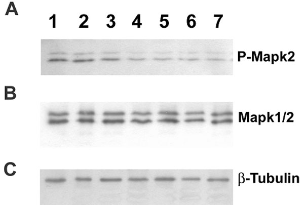
Figure 3. Mitogen-activated protein kinase expression studies in postnatal mouse retina
Postnatal mice retina obtained at: Lane1, (PN) day 0; lane 2, PN 1; lane 3, PN 2; lane 4, PN 3; lane 5, PN 4; lane 6, PN 5, and lane 7, PN 7 were subjected to immunoblot analysis by probing with specific antibodies: A, anti-Phospho-MAPK2; B, anti-MAPK1/2; and C, anti-βTubulin antibodies. The intensity of the immunolabeled bands is directly proportional to the level of specific protein expressed in the retinal samples. Intensity of b-tubulin was used as internal control to normalize the total protein used for analysis.