![]() Figure 6 of
Yang, Mol Vis 2007;
13:1083-1093.
Figure 6 of
Yang, Mol Vis 2007;
13:1083-1093.
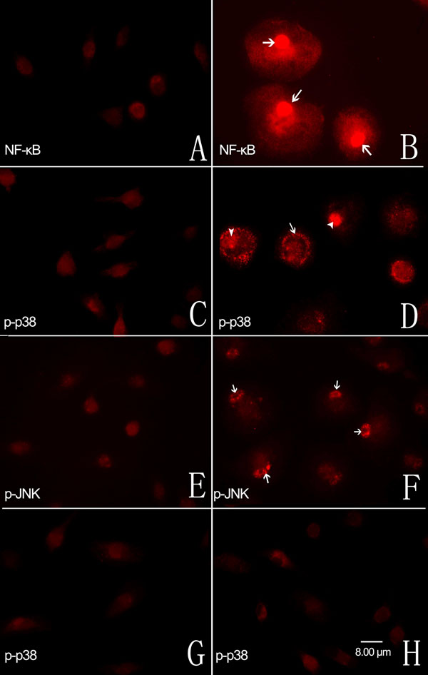
Figure 6. Immunofluorescent localization of the p65 subunit of nuclear factor-κB and the p-p38, and p-JNK mitogen-activated protein kinases in cultured retinal microglial cells
A: Cells expressed very low levels of the p65 subunit of NF-κB without LPS administration. B: Following exposure to LPS for 1 h, p65 subunit labeling was upregulated and translocated into the nuclei (arrow). C: Cells expressed low levels of p-p38 without LPS administration. D: Following exposure to LPS for 1 h, p-p38 expression was upregulated and positive labeing was present in both cytoplasm (arrow) and nuclei (arrowhead). E: Retinal microglial cells expressed low levels of p-JNK without LPS administration. F: Following exposure to LPS for 1 h, the positive labeling of p-JNK was present mainly in nuclei (arrowhead). Pretreatment with minocycline G or sulforaphane H for 1 h followed by LPS administration completely inhibited LPS-induced upregulation of p-p38. All panels have the same scale and the scale bar equal to 8 μm.