![]() Figure 2 of
Yang, Mol Vis 2007;
13:1083-1093.
Figure 2 of
Yang, Mol Vis 2007;
13:1083-1093.
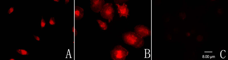
Figure 2. Immunofluorescent characterization of the cultured retinal microglial cells
A: The purified cells were reactive to CD11b, a cell-type-specific marker for microglia. B: After LPS stimulation for 24 h, the CD11b-labeled cells became bigger and rounder, developing the characteristic ameboid shape of activated microglia. C: None of the purified cells showed a positive reaction for GFAP.