![]() Figure 4 of
Yang, Mol Vis 2007;
13:1073-1082.
Figure 4 of
Yang, Mol Vis 2007;
13:1073-1082.
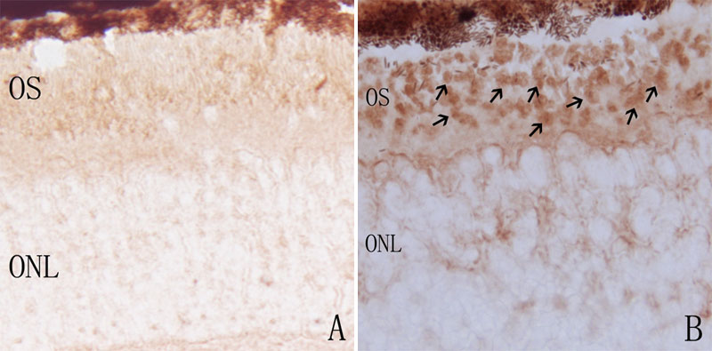
Figure 4. Immunohistochemical labeling of iNOS protein in control and rds mice retinas
A: At P14 in C3B mice, there was no iNOS-positive staining seen. B: At P14 in rds mice, iNOS-positive staining was mainly located at the photoreceptor outer segment (arrows). ONL represents outer nuclear layer; OS represents outer segment. (Magnification x400).