![]() Figure 2 of
Yang, Mol Vis 2007;
13:1073-1082.
Figure 2 of
Yang, Mol Vis 2007;
13:1073-1082.
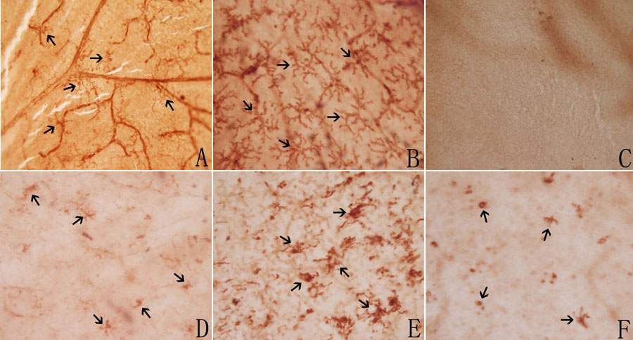
Figure 2. Immunohistochemical labeling of CD11b-positive microglial cells in retinal whole-mounts on control and rds mice
A-C: Normal control C3B mouse retina at P28: (A) In the inner retinal layer, microglia had long, thin processes, located in the peri-vascular region (arrows); (B) In the middle retinal layer, microglia were ramified with short, wide processes. Most cells were vessel-independent (arrows); (C) In the outer nuclear layer (ONL), no microglial cells were identified. D-F: Microglial cells in ONL of rds mice. The cells were ameboid with few stout processes (arrows): (D): At P17, microglial cells were initially present in the ONL; (E): By P28, the number of microglial cells in the ONL had reached their peak; (F): At P180, few microglial cells were scattered in the ONL. (Magnification x400).