![]() Figure 1 of
Yao, Mol Vis 2007;
13:1066-1072.
Figure 1 of
Yao, Mol Vis 2007;
13:1066-1072.
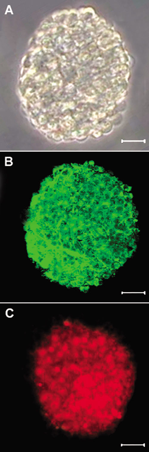
Figure 1. Culture of retinal progenitor cells
A: Phase-contrast micrograph of a neurosphere from passage 3, showing that it is composed of many cells. B: Most cells in the sphere prior to differentiation exhibited Nestin (green). C: Most cells in the sphere prior to differentiation exhibited BrdU (red). Scale bars equal 150 μm.