![]() Figure 1 of
Matsuoka, Mol Vis 2007;
13:1058-1065.
Figure 1 of
Matsuoka, Mol Vis 2007;
13:1058-1065.
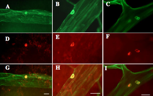
Figure 1. Adherent leukocytes in the retinal vasculature
Confocal fluorescence photographs of retinal vasculature and adherent leukocytes in Sprague-Dawley (SD) rats as control (A, D, G), streptozotocin-induced diabetic rats (STZ), which were spontaneously diabetic Torii (SDT) rats: (B, E, H), and SDT rats (C, F, I). The retinal vasculature and adherent leukocytes were stained green by FITC-conjugated concanavalin A lectin (A, B, C), and the adherent leukocytes were labeled with phycoerythrin-conjugated anti-rat CD45 antibody (D, E, F). The adherent leukocytes appear yellow in the merges images (G, H, I). Scale bar equals 20 μm. J shows the Index of retinal leukostasis. The adherent leukocytes were significantly increased in STZ rats compared to that of control SD rats in short term experiments (control vs STZ, p<0.05), and in long term experiments (p<0.05). SDT rats in short term experiments (p<0.05), and in long term experiments (p<0.05) also showed significantly increased adherent leukocytes compared to that of controls. However, SDT in long term experiments show significantly lower levels of leukostasis compared to that of STZ rats. In addition, the adherent leukocytes were significantly increased with duration of diabetes in STZ rats (p<0.05), but SDT rats did not show the increase with duration of diabetes. The data were analyzed by ANOVA. Asterisk equals p<0.05. DM represents diabetes mellitus. Eight eyes are used in each group at each study period.