![]() Figure 3 of
Gu, Mol Vis 2007;
13:1045-1057.
Figure 3 of
Gu, Mol Vis 2007;
13:1045-1057.
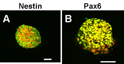
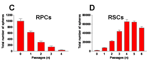
Figure 3. Subsphere formation and gene expression changes with passage
A, B: subcultured retinal stem cell (RSC) spheres express the undifferentiated retinal cell markers nestin (A) and Pax6 (B) by immunohistochemistry (IHC). Scale bars represents 100 μm. Nuclei were stained with propidium iodide. C, D: dissociated retinal progenitor cell (RPC) and RSC spheres generated secondary spheres when grown in suspension. Data are expressed as mean±SD from three independent experiments. E, F: RT-PCR analysis of RNA from RPC (E) and RSC (F) spheres at different passages. M indicates molecular weight marker lane. (-) indicates PCR amplification using cDNA synthesis reactions without reverse transcriptase. β-actin was used as internal control.