![]() Figure 2 of
Gu, Mol Vis 2007;
13:1045-1057.
Figure 2 of
Gu, Mol Vis 2007;
13:1045-1057.
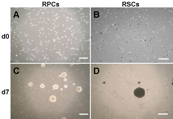
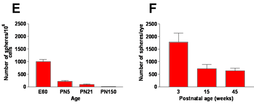
Figure 2. Primary sphere formation in serum-free medium in vitro
A: single cells from primary cultures of dissociated E60 retina-derived cells and B: of 3 week old ciliary epithelium (CE)-derived cells at day 0. Dissociated CE cultures comprise pigmented and non-pigmented cells (B). C, D: primary sphere colonies at day 7 after plating, showing pigmented spheres in CE-derived RSC cultures (D). Scale bars represent 200 μm. E, F: number of primary spheres formed from retina (E) and CE-derived (F) primary cultures at different ages. Three week old CE cultures generated more primary spheres than those from 15 and 45 week old pigs (F). Data are expressed as mean±SD from three independent experiments.