![]() Figure 1 of
Gu, Mol Vis 2007;
13:1045-1057.
Figure 1 of
Gu, Mol Vis 2007;
13:1045-1057.
Figure 1. Identification of retinal progenitor cells and retinal stem cells in the developing and mature retina and ciliary epithelium
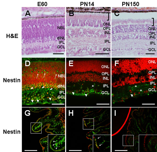
A-C: light microphotographs of H&E stained 15 μm retina cryosections. At PN14 (B) retinal histology was characteristic of the mature (PN150) retina (C) with each of the nuclear and plexiform layers readily identifiable. Mature outer segments of the photoreceptors (brackets) were already evident in the PN14 retina. The apparent detachment of the retina in C is a histological artifact. D-F: confocal fluorescent microphotographs of retina and G-I: of ciliary body (CB) cryosections (15 μm) immunostained with anti-nestin antibody (green). At E60 nestin immunoreactivity was observed in the ganglion cell layer (GCL; arrowheads), (developing) inner nuclear layer (dINL; thick arrows), neuroblast layer (NBL; thin arrows), and in the inner plexiform layer (IPL). At PN14 (E) nestin immunostaining was observed in the GCL (arrowheads) and IPL (asterisk). By PN150 (F) nestin immunoreactivity was observed in fibers in the GCL and IPL (asterisks), and in sparse cells in the GCL (arrowhead). At both E60 (G) and PN14 (H) nestin immunoreactivity was observed in cells distributed within the CB epithelium (thin arrows in insets at bottom). A higher percentage of cells with intense nestin immunostaining were observed in the E60 CB (G). Nestin immunoreactivity was not detected in the PN150 CB (I). Insets in G-I represent higher magnification images of the marked areas. Labeled stromal cells in G most likely represent migrating precursors of neural crest origin. The red line in I corresponds to propidium iodide (PI) staining in the adjacent lens. Nuclei were counterstained with PI (red). Scale bars: A, G-I, 50 μm; D-F, 100, B-C, 200 μm. ONL represents outer nuclear layer, OPL represents outer plexiform layer.