![]() Figure 1 of
Riveiro-Alvarez, Mol Vis 2007;
13:96-101.
Figure 1 of
Riveiro-Alvarez, Mol Vis 2007;
13:96-101.
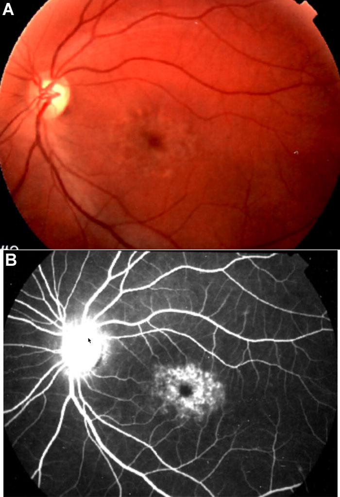
Figure 1. Ophthalmological findings
A: Fundus photograph of the patient's left eye shows macular yellow flecks characteristic of Stargardt macular dystrophy. B: Angiofluoresceingraphy (AFG) shows silent choroid and atrophic retinal pigment epithelium at the macular area.