![]() Figure 3 of
Fuse, Mol Vis 2007;
13:1005-1009.
Figure 3 of
Fuse, Mol Vis 2007;
13:1005-1009.
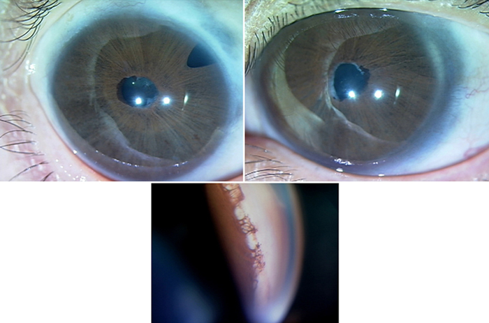
Figure 3. Clinical features of proband in Family 2
Top: The photo shows the eyes of proband affected with ARS. These eyes display megalocornea, posterior embryotoxon, iris hypoplasia, and right iridectomy (post-trabeculectomy). Bottom: Gonioscopic appearance of patients with proband illustrates iridocorneal angle anomaly. This appearance reveals tissue strands extending from peripheral iris to prominent Schwalbe's line and a high insertion of iris into trabecular meshwork.