![]() Figure 2 of
Karring, Mol Vis 2007;
13:997-1004.
Figure 2 of
Karring, Mol Vis 2007;
13:997-1004.
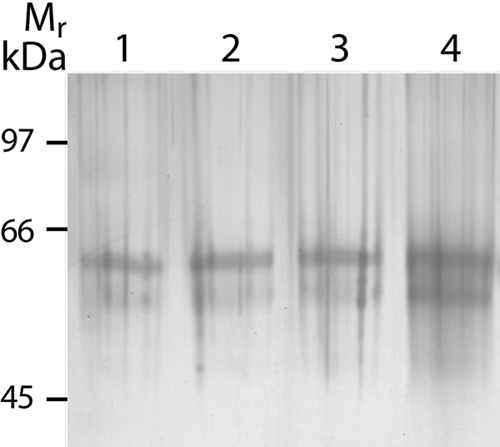
Figure 2. Analysis and identification of the environmental protein contaminants
Silver stained SDS gel of blank and dust samples. Lanes 1-3 show blank samples containing sample buffer and 35 mM DTT. Lane 4 shows proteins extracted from 25 μg of normal environmental dust in sample buffer containing 35 mM DTT. The upper band (about 65 kDa) is keratin-1 and the lower band (60 kDa) is keratin-10.