![]() Figure 4 of
Micera, Mol Vis 2007;
13:981-987.
Figure 4 of
Micera, Mol Vis 2007;
13:981-987.
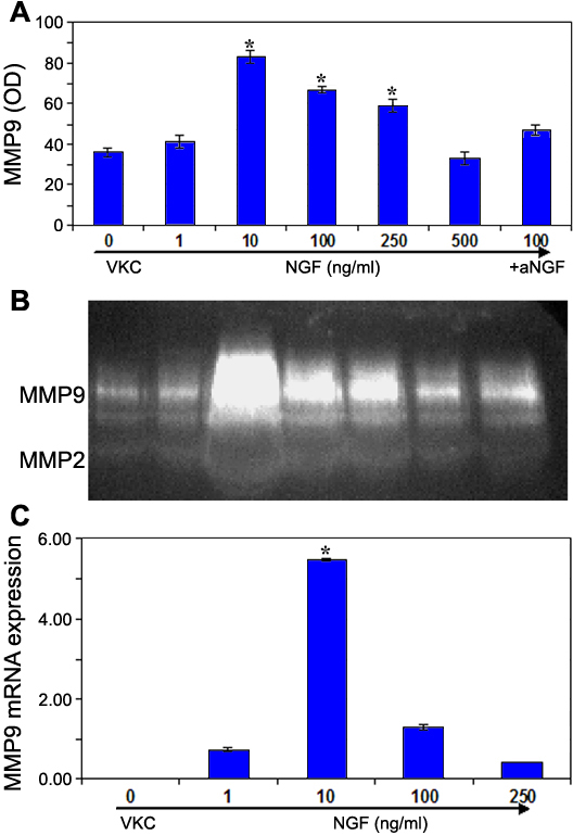
Figure 4. Nerve growth factor increases MMP9 expression and function by vernal keratoconjunctivitis-derived fibroblasts
Conditioned media were collected and processed as described in Methods. A: The histogram shows a significant increase of MMP9 protein expression in VKC-FBs treated with increasing NGF concentrations, according to the densitometric analysis (mean OD±SEM; p<0.05). Data were normalized to GAPDH expression and presented as fold increase with respect to untreated VKC-FBs. B: The functional activity in MMP9 was investigated by SDS-PAGE zymography. From left to right (1-6 lines): 0, 1, 10, 100, 250, 500 ng/ml NGF; line 7, αNGF+100 ng/ml NGF. Panel represents one of three independent gels that gave the same results. C: Relative real-time PCR showed a significant increase of MMP9 mRNA expression in VKC-FBs treated with different concentrations of NGF (p<0.05). Data were normalized to GAPDHmRNA expression and presented as fold increase [11] with respect to untreated VKC-FBs.