![]() Figure 1 of
Micera, Mol Vis 2007;
13:981-987.
Figure 1 of
Micera, Mol Vis 2007;
13:981-987.
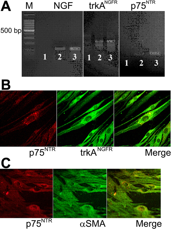
Figure 1. Nerve growth factor, trkANGFR, p75NTR and αSMA expression by vernal keratoconjunctivitis-derived fibroblasts
A: Conventional RT-PCR. showing from left to right, nerve growth factor (NGF; 120 bp), trkANGFR (103 bp); p75NTR (100 bp) amplicons. The lines are as follows: (1) -RT, (2) healthy-FBs, and (3) VKC-FBs. These are representative gels from three independent experiments where equal amounts of cDNA were amplified. B: Confocal microscopy on VKC-FBs showing, from left to right, that VKC-FBs express p75NTR (Cy3, red) and trkANGFR (Cy2, green). p75NTR and trkANGFR colocalized in some cellular compartments (merge; X600/oil immersion). C: Confocal microscopy on VKC-FBs showing, from left to right, that these cells express p75NTR (Cy3, red) and αSMA (Cy2, green). p75NTR and αSMA colocalized in some cellular compartments (merge; X600/oil immersion).