![]() Figure 4 of
Sullivan, Mol Vis 2007;
13:975-980.
Figure 4 of
Sullivan, Mol Vis 2007;
13:975-980.
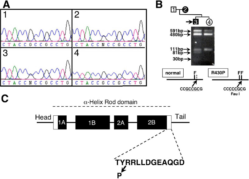
Figure 4. Mutation analysis of the KRT12 gene
A: Bidirectional sequence analysis of the KRT12 gene. The results shown are in the sense direction. KRT12 sequence with in the helix-terminal motif of rod domain 2B in affected family members (2 and 3) showing a G to C transversion at the at the 2nd position of codon 430 that results in an amino acid change from arginine to proline. 1, unaffected father; 2, affected mother; 3, proband and 4, unaffected sister. B: Restriction endonuclease analysis was used to detect R430P mutation. Amplicons of exon 6 were digested with FauI, size fractionated on a 2.5% agarose gel, and visualized under ultraviolet light after staining with ethidium bromide. An additional FauI site (generated by the R430P mutation) converts the 111 bp fragment into 81 and 30 bp fragments. Due to poor enzyme activity, undigested PCR product (591 bp) was observed. The 30 bp fragment was difficult to visualize. C: The domain structure of KRT12 and the mutation position found in MCD family in this study is shown. The rod domain comprised four segments (1A, 1B, 2A, and 2B), represented by filled boxes. The helix-initiation and -termination motif are represented by white boxes. The amino acid sequence for the helix-termination motif is shown.