![]() Figure 3 of
Sullivan, Mol Vis 2007;
13:975-980.
Figure 3 of
Sullivan, Mol Vis 2007;
13:975-980.
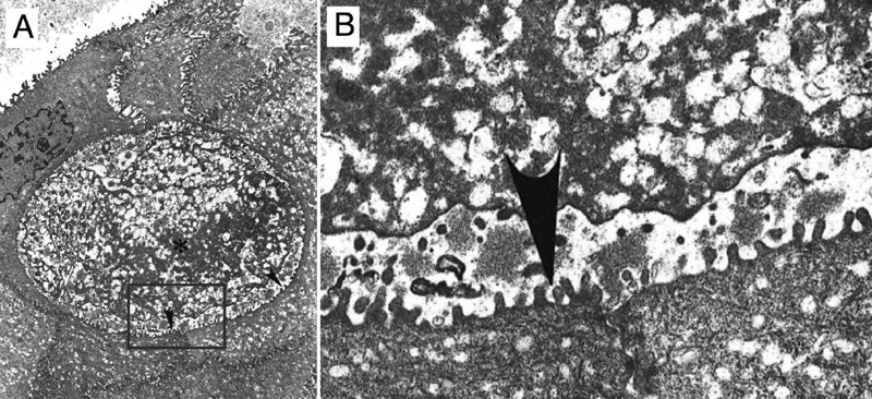
Figure 3. Presence of a "peculiar" electron-dense substance in the intraepithelial cysts of the corneal epithelium sampled from the proband
A: Electron micrograph of corneal epithelium depicting an intraepithelial cyst containing a "peculiar" electron-dense substance (asterisk) intermixed with small vacuoles and electron-dense filamentous material (original magnification 5,400x). B: Higher magnification of A. The cyst was bordered by numerous microvillous processes (arrowheads).