Figure 2 of Sullivan, Mol Vis 2007; 13:975-980.
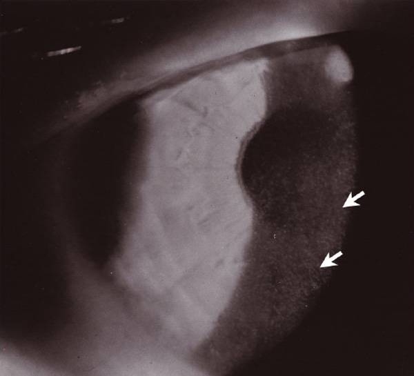
Figure 2. Slit-lamp photograph demonstrating discrete microcysts in the anterior corneal epithelium
Microcysts ranged from clear vesicles to opacified inclusions.