![]() Figure 4 of
Pauli, Mol Vis 2007;
13:962-967.
Figure 4 of
Pauli, Mol Vis 2007;
13:962-967.
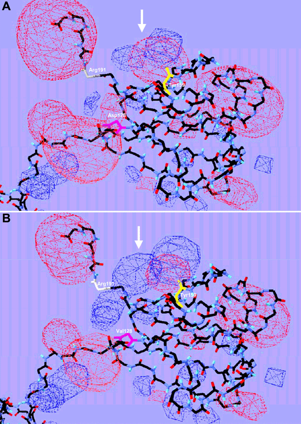
Figure 4. Three-dimensional protein structure
Schematic view of predicted three-dimensional protein structures for CRYBB2WT (A) and CRYBB2D128V (B), without side chains. The electrostatic potential is displayed in red (negative potential) and blue (positive potential) clouds. The alteration from a negative to a positive potential is indicated by white arrows. Exchanged amino acids, Asp and Val, at position 128 are marked in pink. Amino acids involved into H-bond building are shown in white (Arg, 191) and yellow (Tyr, 159).