![]() Figure 7 of
Martinez-Navarrete, Mol Vis 2007;
13:949-961.
Figure 7 of
Martinez-Navarrete, Mol Vis 2007;
13:949-961.
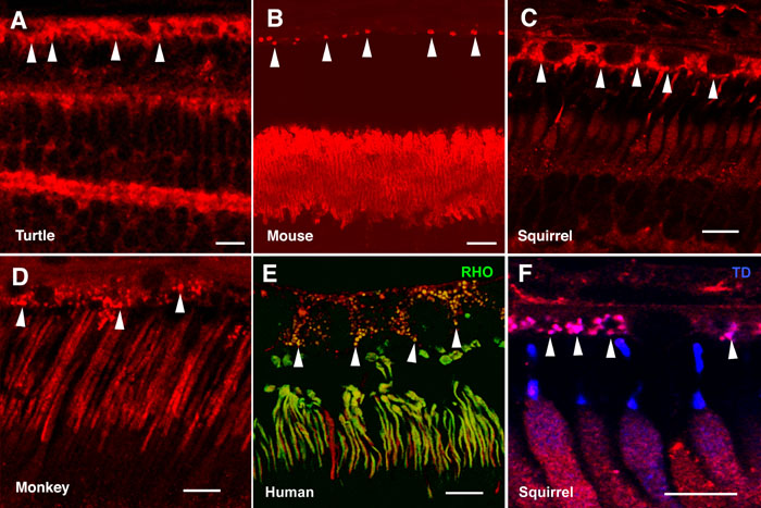
Figure 7. Expression of α-synuclein in retinal epithelial cells
Shown are immunolabeled retina sections of retinal pigment epithelium and photoreceptor outer segment layers for (A) turtle, (B) Swiss mouse, (C, F) squirrel, (D) monkey, and (E) human retinas. α-Synuclein was immunostained red in all panels, rhodopsin (RHO) stained green in panel E; and cone transducin (TD) stained blue in panel F. Arrowheads point to phagosome-like structures in the apical region of retinal epithelial cells. Each bars equals 10 μm.