![]() Figure 6 of
Martinez-Navarrete, Mol Vis 2007;
13:949-961.
Figure 6 of
Martinez-Navarrete, Mol Vis 2007;
13:949-961.
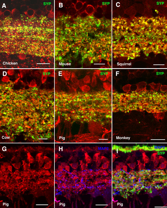
Figure 6. Colocalization between α-synuclein and synaptophysin in the inner plexiform layer
Shown are immunolabeled retina sections of (A) chicken, (B) mouse, (C) squirrel, (D) cow, (E, G-I) pig, and (F) monkey. α-Synuclein immunostained red in all panels, synaptophysin (SYP) stained green in panels A-F and I, and microtubule-associated protein-2 (MAP2) stained blue in panels H and I. Lack of colocalization between α-synuclein and synaptophysin is evident in panel I. Each bar equals 10 μm.