![]() Figure 4 of
Martinez-Navarrete, Mol Vis 2007;
13:949-961.
Figure 4 of
Martinez-Navarrete, Mol Vis 2007;
13:949-961.
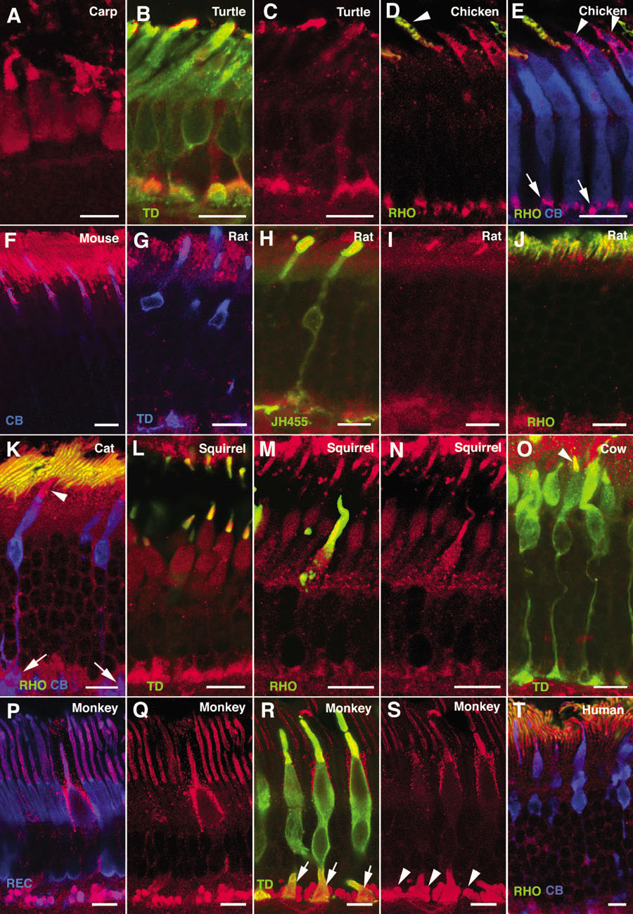
Figure 4. Expression of α-synuclein in photoreceptor cells
Shown are immunolabeled views of (A) carp, (B, C) turtle, (D, E) chicken, (F) mouse, (G-J) rat, (K) cat, (L-N) squirrel, (O) cow, (P-S) monkey, and (T) human photoreceptors. α-Synuclein immunostained red in all panels. Single, double, or triple labelings for different photoreceptor markers are shown. Cone transducin (TD) stained green in panels B, L, O, and R, and stained blue in panel G. Rhodopsin (RHO) stained green in panels D, E, J, K, M, and T, and S-cone opsin (JH455) stained green in panel H. Calbindin (CB) stained blue in panels E, F, K, and T, as did recoverin (REC) in P. Arrowheads point to outer segments of rods (panel D) and cones (panels E, K, O). Arrows mark cone pedicles (panels E, K, R), while arrowheads point to rod spherules (panel S). Each bars equals 10 μm.