![]() Figure 3 of
Martinez-Navarrete, Mol Vis 2007;
13:949-961.
Figure 3 of
Martinez-Navarrete, Mol Vis 2007;
13:949-961.
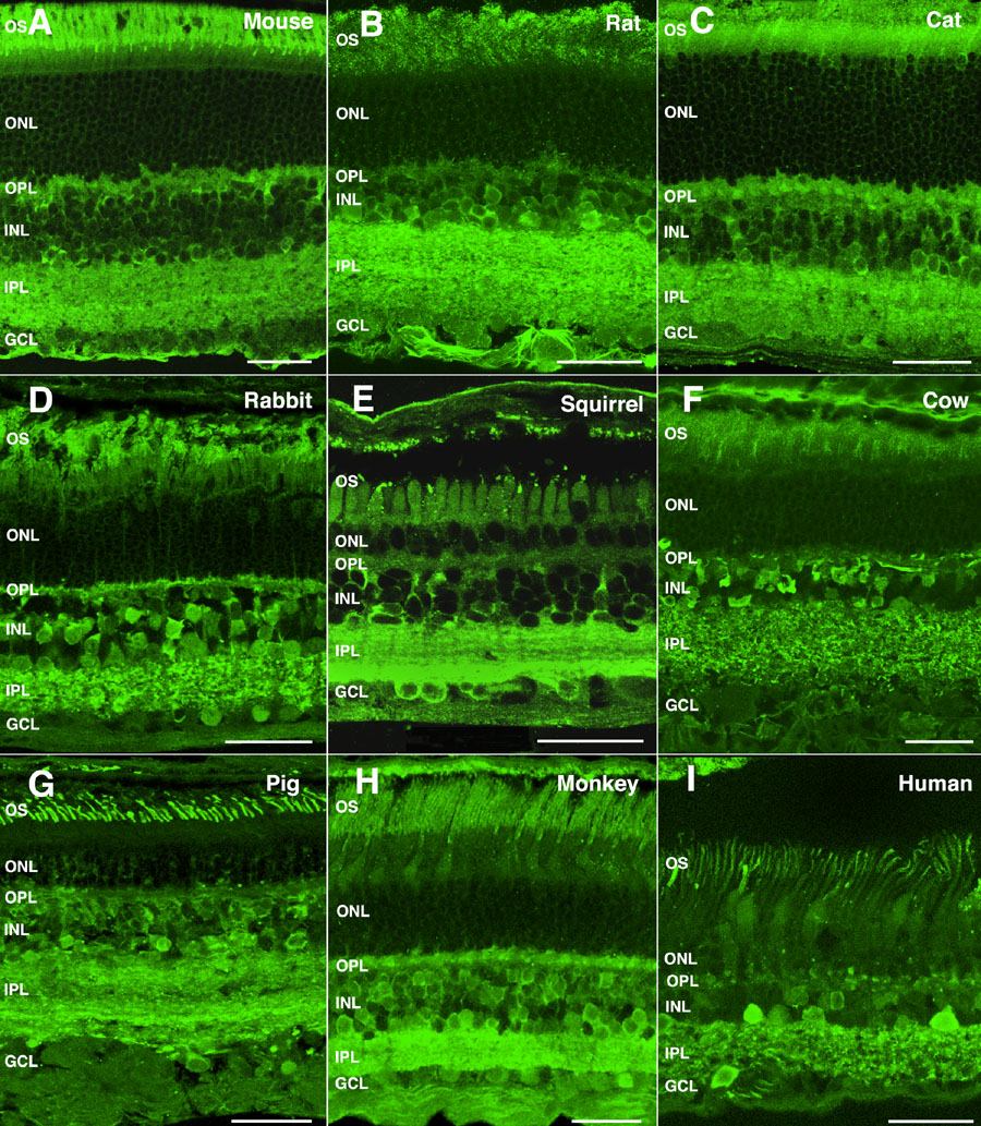
Figure 3. α-Synuclein immunoreactivity pattern in the retina of mammalian vertebrates
Shown are retinal sections immunolabeled for α-synuclein from (A) mouse, (B) rat, (C) cat, (D) rabbit, (E) squirrel, (F) cow, (G) pig, (H) monkey, and (I) human. The following abbreviations were used: outer nuclear layer (ONL), ganglion cell layer (GCL), inner nuclear layer (INL), outer plexiform layer (OPL), inner plexiform layer (IPL), and outer segments (OS). Each bars equals 40 μm.