![]() Figure 2 of
Martinez-Navarrete, Mol Vis 2007;
13:949-961.
Figure 2 of
Martinez-Navarrete, Mol Vis 2007;
13:949-961.
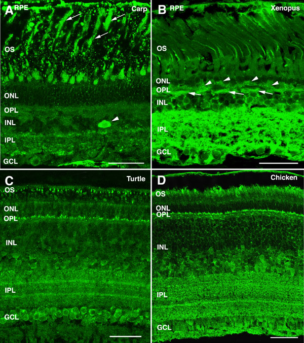
Figure 2. α-Synuclein immunoreactivity pattern in the retina of non-mammalian vertebrates
Shown are retinal sections from (A) carp, (B) Xenopus, (C) turtle, and (D) chicken immunolabeled for α-synuclein. Arrows point to rod outer segments in panel A and to horizontal cells in panel B. Arrowheads point to amacrine cells in panel A and to photoreceptor axon terminals in panel B. The following abbreviations were used: outer nuclear layer (ONL), ganglion cell layer (GCL), inner nuclear layer (INL), outer plexiform layer (OPL), inner plexiform layer (IPL), retinal pigment epithelium (RPE), and outer segments (OS). Each bars equals 40 μm.