![]() Figure 3 of
Zaghloul, Mol Vis 2007;
13:86-95.
Figure 3 of
Zaghloul, Mol Vis 2007;
13:86-95.
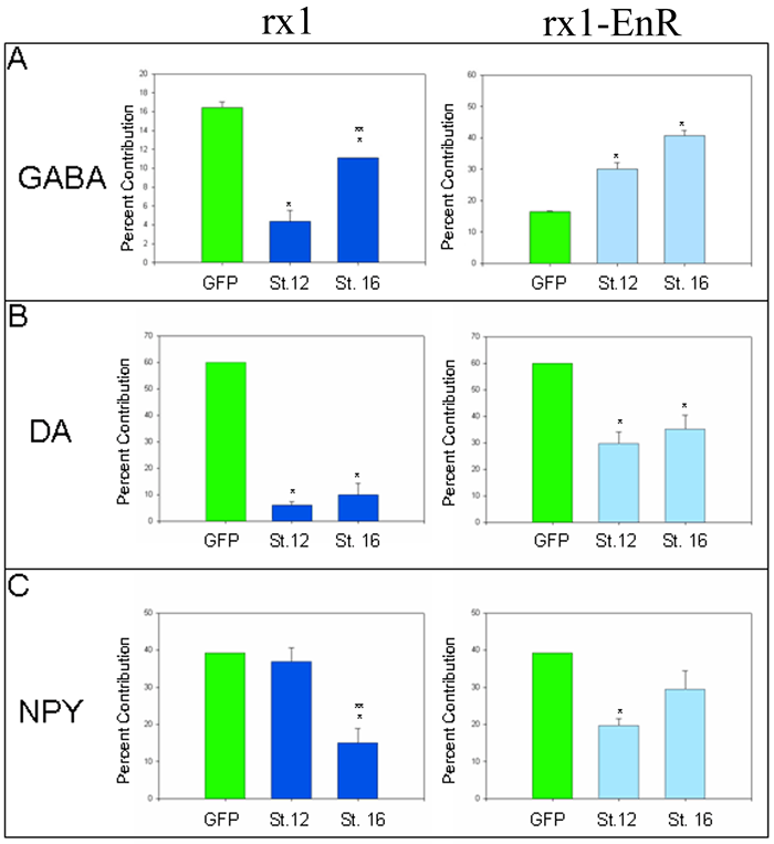
Figure 3. Altered Rx1 levels/activity affect all three amacrine cell subtypes
rx1 gain-of-function (dark blue bars) and loss-of function (rx1-EnR; light blue bars) were induced at early (St. 12) or late (St. 16) eye field stages. The percent contribution that the injected blastomere lineage made to the total number of the subtype was determined for: (A) GABA, (B) DA and (C) NPY amacrine cells. Bars indicate SEM. A single asterisk (*) indicates a significant difference (p<0.05) compared to gfp mRNA-injected control embryos that also were treated with dexamethasone (green bars). A double asterisk (**) over a stage 16 bar indicates a significant difference (p<0.05) compared to stage 12 induction data. All samples passed the equal variance test. rx1 gain-of-function at both eye field stages caused a significant reduction of GABA and DA amacrine cells, but the GABA reduction was significantly less at stage 16. In contrast, NPY amacrine cells were reduced only at the late stage. rx1 loss-of-function increased GABA cell production and reduced DA amacrine cell production equivalently at both eye field stages; it significantly decreased NPY amacrine cells only at stage 12.
