![]() Figure 2 of
Zaghloul, Mol Vis 2007;
13:86-95.
Figure 2 of
Zaghloul, Mol Vis 2007;
13:86-95.
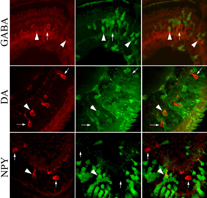
Figure 2. Labeling of amacrine cell subtypes
Sections of retina were labeled with antibodies to distinguish amacrine subtypes (red cells); those descended from the injected blastomere express GFP (green cells). Top row: Large numbers of amacrine cells express GABA (left panel). Large arrowheads indicate two GABA amacrine cells descended from D1.1.1 blastomere (green in middle panel and double-labeled in merged right panel). Small arrow indicates a GABA amacrine cell that is not GFP-labeled. Middle row: Dopamine (DA) amacrine cells are less abundant (left panel). Large arrowhead indicates a DA amacrine cell descended from D1.1.1 blastomere (green in middle panel and double-labeled in merged right panel). Small arrows indicate two DA amacrine cells that are not GFP-labeled. Bottom row: NPY amacrine cells also are less abundant (left panel). Large arrowhead indicates a NPY amacrine cell descended from D1.1.1 blastomere (green in middle panel and double-labeled in merged right panel). Small arrows indicate two NPY amacrine cells that are not GFP-labeled. Each image was collected with 40x oil lens, zoom set at 1.9, in a 1024x1024 pixel field, and pixel size equal to 0.12 μm.