![]() Figure 6 of
Qi, Mol Vis 2007;
13:1-11.
Figure 6 of
Qi, Mol Vis 2007;
13:1-11.
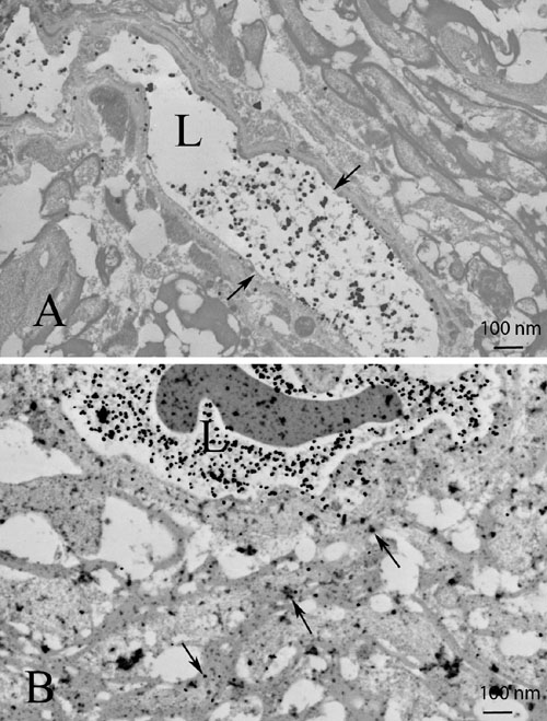
Figure 6. Restoration of blood-brain barrier integrity
Transmission electron micrographs show that the combined effects of catalase and ECSOD markedly decreased extravasation of serum albumin labeled by immunogold (arrows) from the vessel lumen into the perivascular space (A), relative to perivascular accumulation of labeled serum albumin in the unprotected optic nerve (B). The barplot shows mean extravasated albumin immunogold counts in the retrobulbar optic nerves protected by ECSOD and catalase (ECSOD OD), catalase (Wt OD), ECSOD (ECSOD OS) or unprotected EAE (Wt OS; C). Barplot (D) illustrates the decrease in extravasated albumin immunogold in nerves treated with ECSOD and catalase, catalase or ECSOD relative to the unprotected nerves. Asterisk (*) represents p<0.05, double asterisks (**) represents p<0.01, L represents lumen.