![]() Figure 2 of
Qi, Mol Vis 2007;
13:1-11.
Figure 2 of
Qi, Mol Vis 2007;
13:1-11.
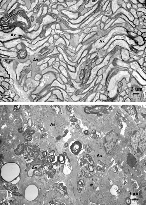
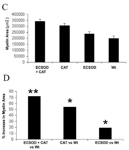
Figure 2. Suppression of demyelination
Representative transmission electron micrographs of the retrobulbar optic nerve show many normal fibers and substantially less demyelinated and thinly myelinated axons following rAAV-catalase inoculation of the right eyes of transgenic ECSOD mice (A), relative to the unprotected left eyes of wild-type littermates in whom marked fiber loss, naked axons and those with thin sheaths of myelin were prominent ultrastructural findings (B). The barplot shows mean myelin areas of the retrobulbar optic nerve protected by both ECSOD and catalase (ECSOD OD), catalase (Wt OD), ECSOD (ECSOD OS) and unprotected EAE (Wt OS; C). Barplot (D) illustrates the preservation of myelin induced by ECSOD and catalase, catalase or ECSOD relative to unprotected nerves. Asterisk (*) represents p<0.05, double asterisks (**) represents p<0.01, Ax represents axon, As represents astrocyte process.