![]() Figure 1 of
Qi, Mol Vis 2007;
13:1-11.
Figure 1 of
Qi, Mol Vis 2007;
13:1-11.
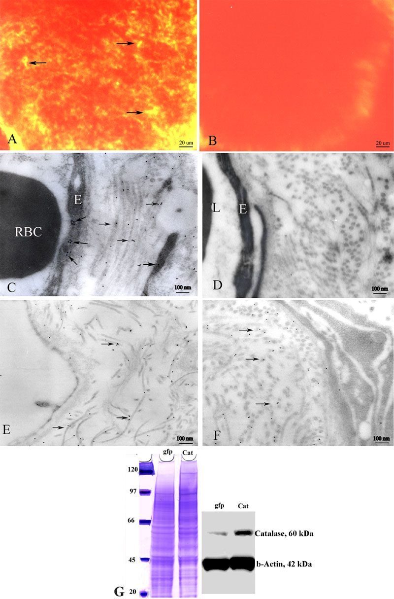
Figure 1. Expression of extracellular superoxide dismutase and catalase
Immunofluorescence micrographs show expression of the human ECSOD (arrows) in the optic nerve of a representative transgenic ECSOD mouse (A), but it is absent in the optic nerve of a wild-type littermate (B). Transmission electron micrograph of the retrobulbar optic nerve of a transgenic ECSOD mouse reveals ECSOD immunogold (arrows) in the perivascular space and endothelia of the optic nerve (C), peripapillary choroid (E) and optic nerve sheath (F). Human ECSOD is absent in wild-type littermates (D). Immunobloting shows increased catalase expression in cultured retinal ganglion cells infected with rAAV containing the gene for human catalase, relative to control RGC-5 cells infected with AAV-GFP (G). E represents endothelial cell. RBC represents red blood cell. L represents lumen, Cat represents catalase, gfp represents green fluorescent protein.