![]() Figure 6 of
Nakazawa, Mol Vis 2006;
12:867-878.
Figure 6 of
Nakazawa, Mol Vis 2006;
12:867-878.
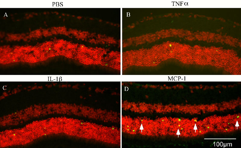
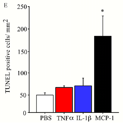
Figure 6. MCP-1 exacerbates photoreceptor cell apoptosis
A-D: Representative photomicrographs of merged images of TUNEL (green) and nuclear staining with propidium iodide (red) from rat retinal sections receiving subretinal injection of PBS (A), TNF-α (B), IL-1β (C), or MCP-1 (D). Retinal sections were prepared at 24 h post-injection. Arrow points to TUNEL-positive cells. E: Bar chart indicates counts of TUNEL-positive cells per mm2 of retinal section (n=7). Data presented are means; error bars represent the SEM. Asterisk (*) indicates p<0.05 compared to PBS treated group.