![]() Figure 4 of
Nakazawa, Mol Vis 2006;
12:867-878.
Figure 4 of
Nakazawa, Mol Vis 2006;
12:867-878.
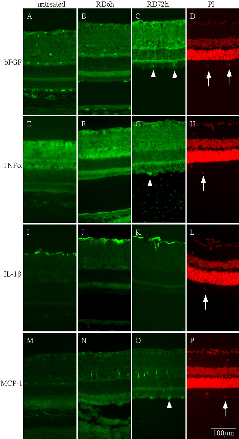
Figure 4. Immunohistochemical analysis of cytokines after RD
Representative photomicrographs of retinal sections labeled with primary antibodies against bFGF (A-D), TNF-α (E-H), IL-1β (I-L), and MCP-1 (M-P) or nuclear staining dye propidium iodide (PI: D,H,L,P). The retinal sections were derived from the control eye (A,E,I,M) or those at 6 (RD 6 h; B,F,J,N) or 72 h after RD (RD 72 h; C,G,K,O; n=4 each). Arrows point to monocytes, and arrowheads indicate the positive immunoreactivity of bFGF (C), TNF-α (G), or MCP-1 (O). Note that immunoreactivity of bFGF appeared in retinal sections at 72 h after RD; whereas, the upregulation of TNF-α, IL-1β, and MCP-1 immunoreactivity started by 6 h.