![]() Figure 5 of
Yin, Mol Vis 2006;
12:858-866.
Figure 5 of
Yin, Mol Vis 2006;
12:858-866.
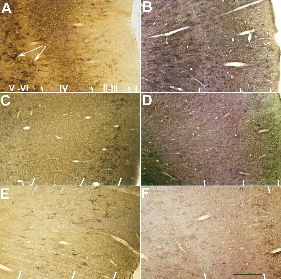
Figure 5. Distribution of cortical Cat-301 labeling affected in strabismic and Y-block cat
Noncounterstained photomicrographs from visual cortex of normal (A), strabismic amblyopic (C), and Y-blocked (E) cats showing laminar distribution of immunoreactivity. The cortical layers, I, II-III, IV, and V-VI are demarcated by indicator lines at the bottom of each photograph. In the normal cat, the distribution of Cat-301 immunopositivity was prominent in 2 bands of layer III-IV and layer V-VI. Note the outlining of somata and proximal dendrites in individual neurons of layer V-VI (white arrows). In the strabismic amblyopic cat, Cat-301-labeled neurons were mainly reduced in layer IV. Only a few Cat-301-labeled cells were seen in upper layers II-III of Y-blocked cat. Counterstained photomicrographs from visual cortex of normal (B), strabismic amblyopic (D), and Y-blocked (F) cats. Scale bar represents 0.5 mm.