![]() Figure 4 of
Yin, Mol Vis 2006;
12:858-866.
Figure 4 of
Yin, Mol Vis 2006;
12:858-866.
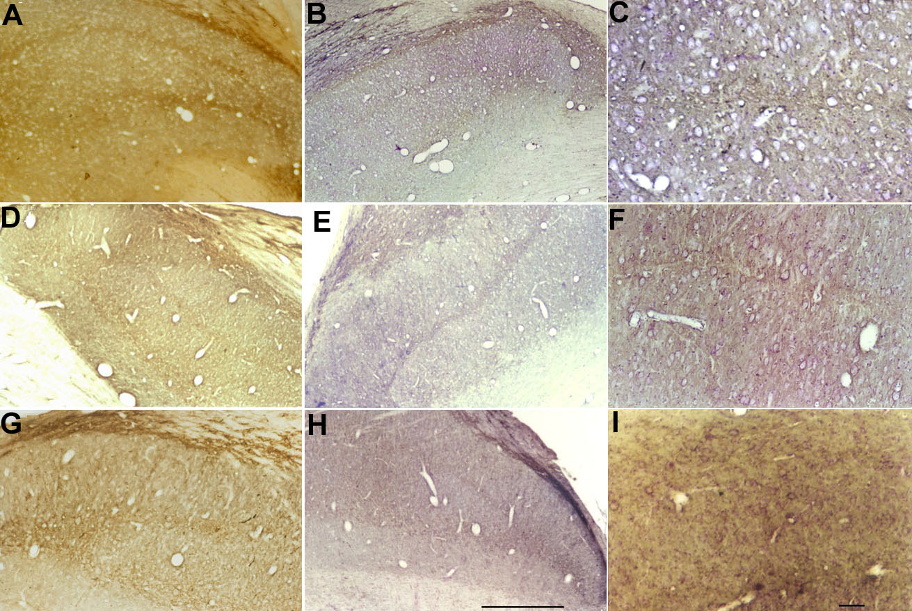
Figure 4. Lower perineuronal staining in strabismic and Y-block lateral geniculate nucleus
Low power magnification micrographs of the LGN of normal (A,B), strabismic (D,E), and Y-block (G,H; scale bar represents 1.0 μm) cats and medium power images (C,F,I; scale bar represents 100 μm). Sections in Panels A, D, and G are not counterstained while B, E, and H are counterstained with cresyl violet. Positive labeling for Cat-301 antibody was found in laminae A, A1, and C laminae, with particular density in interlaminar zones and in the medial interlaminar nucleus. The perigeniculate nucleus that lies dorsal to the LGN also stained intensely. The level of nonsomatic (dendritic and background) labeling is not obviously different between laminae receiving input from the two eyes in any of the conditions, but is reduced for strabismic and Y-block sections compared with normal. At higher magnification, the perineuronal staining is evident in many of the neurons of normal (C), while there is a lower frequency of perineuronal staining in strabismic (F) and Y-block (I).