![]() Figure 1 of
Yin, Mol Vis 2006;
12:858-866.
Figure 1 of
Yin, Mol Vis 2006;
12:858-866.
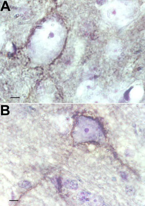
Figure 1. Cat-301 staining of neurons in LGN and cortex
A: Cat-301 antibody binds to the cell membrane, somata, and proximal dendrites of Y-cells in lateral geniculate nucleus (LGN) of normal cat. B: Visual cortical neurons recognized by Cat-301. The antibody staining outlines the cell body, somata, proximal dendrites, and axon of Cat-301-positive cells in visual cortex. Cells were counterstained with cresyl violet (100x objective). Cells were counted only if the cresyl stained nucleolus was visible. Clear examples of labeled and unlabeled neurons are visible in both Panels A and B. Scale bars represent 5 μm.