![]() Figure 6 of
Yamanaka, Mol Vis 2006;
12:841-851.
Figure 6 of
Yamanaka, Mol Vis 2006;
12:841-851.
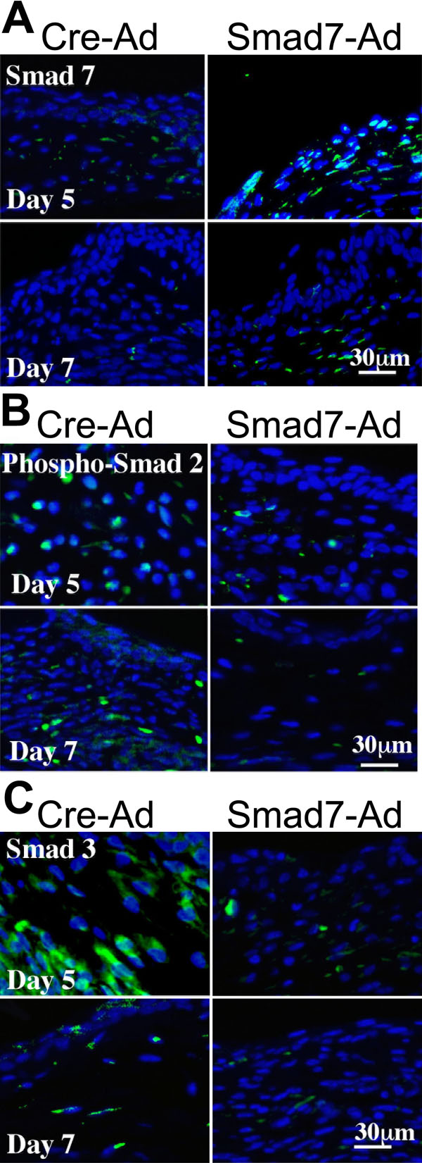
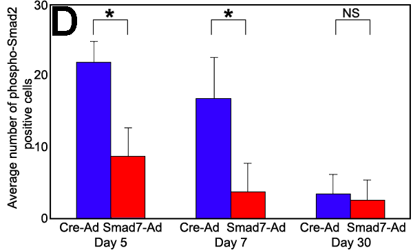
Figure 6.
Immunolocalization of Smad in mechanically-injured mice conjunctiva. Smad7 protein, presumably the translated produce derived from exogenously introduced Smad7 cDNA, was strongly detected in the healing conjunctival epithelium and fibroblasts from day 2 onward, while faint immunoreactivity for endogenous Smad7 was seen in the healing conjunctiva in the control group for up to 10 days (A). At day 5, phosphorylated Smad2 and Smad3 were both detected in the nuclei of conjunctival fibroblasts and epithelial cells in control eyes, whereas in Smad7-Ad treated eyes phospho-Smad2 was not detected and Smad3 was seen only faintly in the cytoplasm (B,C). At and after day 10, a few of the nuclei were positive for both Smad2 and Smad3 in the control, but negative in Smad7-Ad treated specimens. The numbers of phospho-Smad2-positive fibroblasts were significantly lower in Smad7-Ad treated group than in Cre-Ad control group, while there was no significant difference on day 30 (D). The asterisks in Panel D indicates a p<0.01; NS represents nonsignificant.