![]() Figure 5 of
Yamanaka, Mol Vis 2006;
12:841-851.
Figure 5 of
Yamanaka, Mol Vis 2006;
12:841-851.
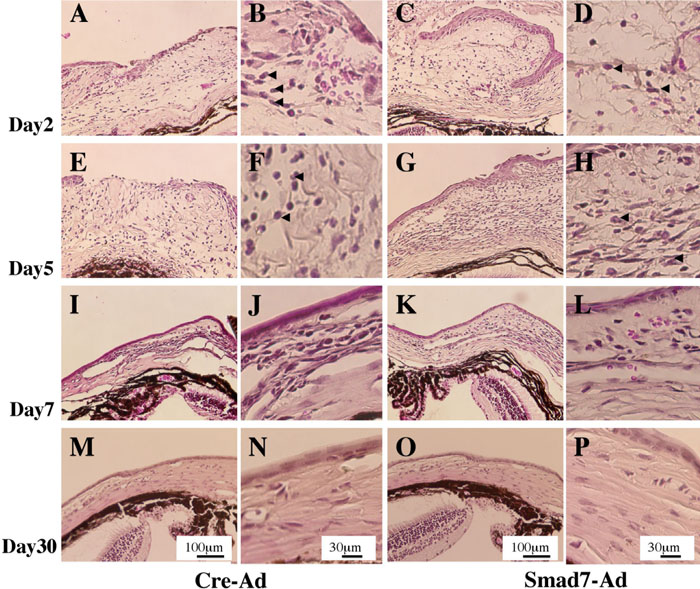
Figure 5.
H&E histology in a mechanically injured mice conjunctiva. Smad7 gene introduction seemed to suppress the degree of conjunctival edema and cell population compared to control (A-P). The conjunctival epithelium defect sealed as early as day 5 in the Smad7-Ad group (G,H), whereas the epithelial defect was not closed at day 5 in the Control group (E,F). On day 7, the epithelial defect was closed in both groups (I-L). On day 30, however, H&E histology of both groups of specimens exhibited similar findings: reduced cell population and reduction of the thickness of subconjunctival matrix (M-P). Polymorphonuclear leukocytes were seen in subconjunctival tissue at day 2-5, but there seemed to be no difference of the distribution of this cell type. The scale bar represents 100 μm for A,C,E,G,I,K,M,O and 30 μm for B,D,F,H,J,L,N,P. Arrowheads point to polymorphonuclear leukocytes.