![]() Figure 2 of
Park, Mol Vis 2006;
12:832-840.
Figure 2 of
Park, Mol Vis 2006;
12:832-840.
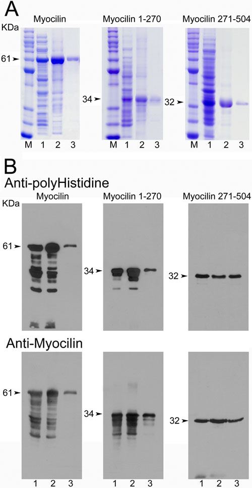
Figure 2.
Purification of full length and truncated myocilins. Samples from different stages of the purification process were resolved on 12% SDS-PAGE gels. Lane 1, bacterial cell lysate; lane 2, solubilized inclusion bodies; and lane 3, nickel-purified proteins. The gels were either stained with Coomassie blue (A) or electroblotted for western analyses (B). The amount of protein samples used for Panel B was 1/100 of that used for Panel A. The blots were probed with anti-polyHistidine (upper panel in B) and anti-myocilin (lower panel in B) specific to either NH2-terminus (for myocilin and myocilin 1-270) or COOH-terminus (for myocilin 271-504). Molecular weights (kDa) of the purified proteins (arrowheads) are shown. The size of marker proteins (M) shown in Coomassie staining from the top is 120, 100, 90, 80, 70, 60, 50, 40, 30, 25, and 20 kDa, respectively.