![]() Figure 2 of
Santhiya, Mol Vis 2006;
12:768-773.
Figure 2 of
Santhiya, Mol Vis 2006;
12:768-773.
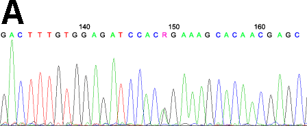
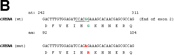
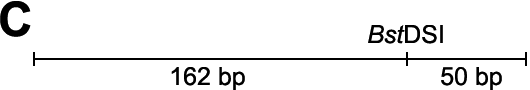
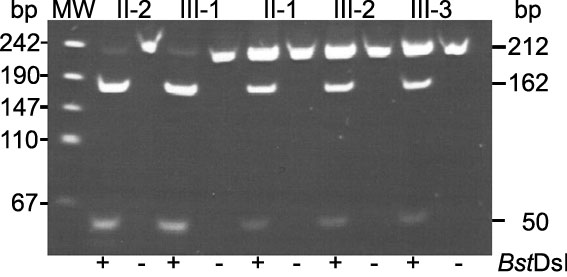
Figure 2.
The CRYAA mutation in family CCE20. A: A partial fragment of the second exon of the CRYAA gene is given at sequence position 149 (position 291 in CRYAA exon 2, counting the A of the ATG start codon as number 1), the heterozygous situation of the proband is obvious (red arrow). B: A partial fragment of the corresponding genomic sequence of the CRYAA gene at the end of exon 2 is given. The mutated sequence is shown. The underlined bases (CCACGG) define a BstDSI restriction site, which is destroyed by the mutation. The wild-type base and amino acid are marked in green, and the mutated forms in red. C: A schematic overview of exon 2 of the CRYAA gene is given indicating the position of the BstDSI restriction site leading to two fragments of 162 and 50 bp. The fragment of 212 bp reflects the mutated form. Exon 2 of the CRYAA gene was amplified in family CCE20 and digested by BstDSI, The fragments were analyzed on an agarose gel. The larger fragment of 212 bp can be observed in the affected family members only. The two smaller fragments are present in all family members (II-2, healthy father; III-1, healthy sister; II-1, affected mother; III-2, affected proband; III-3, affected sib).