![]() Figure 4 of
Pang, Mol Vis 2006;
12:756-767.
Figure 4 of
Pang, Mol Vis 2006;
12:756-767.
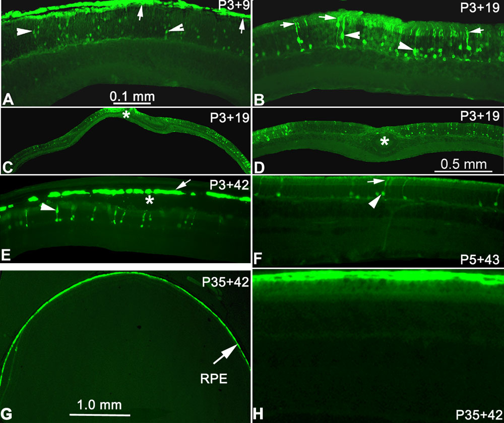
Figure 4. GFP expression in retinal cells after subretinal injection in normal mice
The entire RPE layer expressed GFP (arrows) in a section prepared from an eyecup from a mouse injected on P3 and sacrificed 9 days later (A, P3+9) with relatively weak GFP expression in photoreceptor cells (arrowheads). Panel B shows a section from a retinal whole mount prepared at P3+19 showed GFP expression not only in the photoreceptor nuclei (arrowheads) but also in the inner segments and outer segments of photoreceptor cells (arrows). Note that the RPE layer is not present in this example and that the second arrow from the left of the picture points to a cone photoreceptor that can be identified by the location of the nucleus (directly below the inner segment region) and the expanded synaptic terminal of the cell. Lower magnification of the same section in B showed that a large portion of photoreceptor cells were transduced (C,D; asterisk (*) represents injection site). GFP expression was maintained 6 weeks after injection (E, P3+42; F, P5+43). Note the artificial retinal detachment (asterisk), which was probably related to the subretinal injection (E). GFP expression was detected only in RPE from sections of an eyecup at 3 weeks following trans-corneal subretinal injection at P35 (G,H, P35+42). Panels A,B,E,F,H have the same magnification (the scale bar represents 0.1 mm), and C,G have the same magnification (the scale bar represents 1.0 mm).