![]() Figure 3 of
Pang, Mol Vis 2006;
12:756-767.
Figure 3 of
Pang, Mol Vis 2006;
12:756-767.
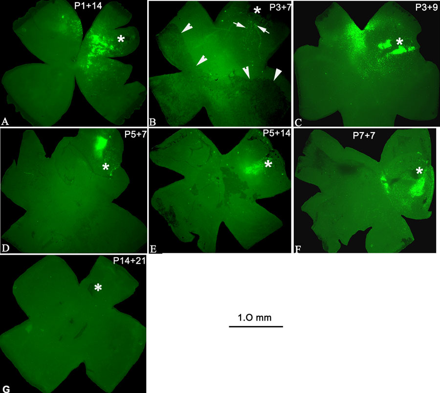
Figure 3. GFP expression in retinal whole mounts from rd mice following subretinal injections
Light micrographs of GFP expression in retinal whole mounts following subretinal injection in rd mice at various ages and sacrificed 1-6 weeks later. GFP expression was intense and covered about 1/4 of the retina two weeks following injection at P1 (and sacrificed 14 days later, A, P1+14). The earliest detectable GFP expression (arrows) was limited to the injection site one week following injection at P3 (B, P3+7). The nonpigmented area superior to arrowheads indicates a retinal detachment due to the subretinal injection. Central bright area is due to autofluorescence. Another mouse injected on P3 and sacrificed 9 days later (P3+9) showed increased fluorescence covering about 1/4 of the retina (C). GFP expression was evident only around the injection site at 1 week after injection at P5 (D, P5+7). GFP expression could also be observed by the second week after injection at P5 (E, P5+14). More intense GFP fluorescence was observed 7 days after injection at P7 (F, P7+7) compared with that injected at younger ages. No GFP expression was detected in the retina 3 weeks following injection at P14 (G, P14+21). Asterisk (*) represents injection site. The scale bar represents 1.0 mm.