![]() Figure 4 of
Augusteyn, Mol Vis 2006;
12:740-747.
Figure 4 of
Augusteyn, Mol Vis 2006;
12:740-747.
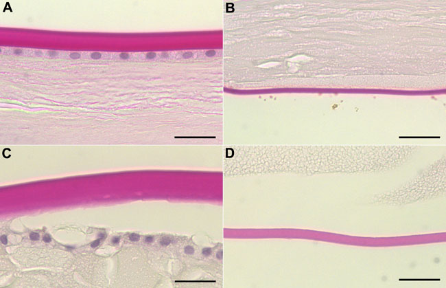
Figure 4.
Light microscopy of the anterior (A) and posterior (B) regions of a 74-year-old human lens without capsular separation and the anterior (C) and posterior (D) regions of a 78-year-old human lens with capsular separation. The bar represents 25 μm. The capsule is stained red and the nuclei stained blue.