![]() Figure 7 of
Pendergrass, Mol Vis 2006;
12:712-724.
Figure 7 of
Pendergrass, Mol Vis 2006;
12:712-724.
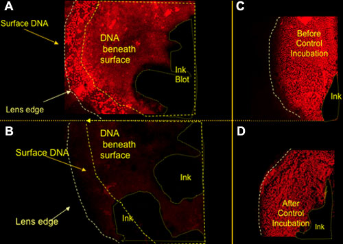
Figure 7.
DNAse I susceptibility of cortical DNA fluorescence in old cataractous lenses. A: Pre-DNAse images of the anterior of an old rat lenses fixed and stained for DNA with PI. The frame has been focused 100 μm below the anterior surface so that both surface and cortical (necrotic inclusions) DNA can be seen (DNA below surface is marked by dashed lines). B: The same lens is shown after DNase 1 digestion for 30 min. C,D: The anterior view of a control young lens before (C) and after (D) control incubation in buffer without DNase 1. The inkblots were used to locate the same areas on the surface of the lens before and following DNAse digestion.